When treating severe burns and trauma, skin regeneration can be a matter of life or death. Extensive burns are usually treated by transplanting a thin layer of epidermis, the top layer of skin, from elsewhere on the body. However, this method not only leaves large scars, it also does not restore the skin to its original functional state. Unless the dermis, the layer below the epidermis, which contains blood vessels and nerves, is regenerated, it cannot be considered normal living skin.
Now, work by Swedish researchers may have brought medicine closer to being able to regenerate living skin. They have developed two types of 3D bioprinting techniques to artificially generate thick skin that is vascularized, meaning it contains blood vessels. One technique produces skin that is packed with cells, and the other produces arbitrarily shaped blood vessels in the tissue. The two technologies take different approaches to the same challenge. The approaches have been outlined in two studies published in the journal Advanced Healthcare Materials.
“The dermis is so complicated that we can’t grow it in a lab. We don’t even know what all its components are,” said Johan Junker, an associate professor at Linköping University and specialist in plastic surgery who lead this work, in a statement. “That’s why we, and many others, think that we could possibly transplant the building blocks and then let the body make the dermis itself.”
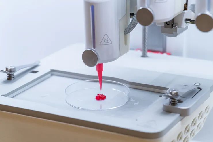
The Linköping team using a 3D bioprinter.
Photograph: Magnus Johansson/Linköping University
Junker and his team designed a bio-ink called “μInk” in which fibroblasts—cells that produce dermal components such as collagen, elastin, and hyaluronic acid—are cultured on the surface of small spongy gelatin grains and encased in a hyaluronic acid gel. By building up this ink three-dimensionally using a 3D printer, they were able to create a skin structure filled with high-density cells at will.
In a transplantation experiment using mice, the researchers confirmed that living cells grew inside tissue fragments made from this ink, secreting collagen and rebuilding the components of the dermis. New blood vessels also grew inside the graft, indicating that the conditions for long-term tissue fixation were met.
Blood vessels play an extremely important role in the construction of artificial tissues. No matter how many cells are cultured to create a tissue model, without blood vessels, oxygen and nutrients cannot be carried evenly to all cells. And without blood vessels, as the tissue structure grows, the cells in the center of the tissue die.
The research team has also created a technology called REFRESH (Rerouting of Free-Floating Suspended Hydrogel Filaments), which enables the flexible construction of blood vessels in artificial tissues by printing and arranging threads of hydrogel, a gels that 98 percent water. These threads are much tougher than ordinary gel materials and can maintain their shape even when tied or braided. Moreover, they also have shape-memory properties that allow them to return to their original shape even when crushed.
A hydrogel thread made using the REFRESH technology.
Photograph: Magnus Johansson/Linköping University
Notably, these threads can be disassembled without leaving any trace by the action of a specific enzyme. When the hydrogel threads placed in the tissue disappear, only a long, thin cavity remains in their original place. By using this as a flow channel equivalent to a blood vessel, a network of blood vessels can be freely formed inside artificially created tissue. By integrating these two technologies, it could be possible to incorporate a freely designed network of blood vessels into the thick, cell-filled artificial skin, allowing oxygen and nutrients to reach every nook and cranny.
The researchers also succeeded in constructing a complex 3D network by forming the hydrogel threads into knots or braids. In the future, they hope to combine this with technology to automate such operations, thereby realizing a method to efficiently stretch a network of blood vessels throughout an artificial organ.
There remain many uncertainties in the wound environment, such as how to avoid inflammation and bacterial infection, and careful verification of these techniques will be needed to bridge the gap between these results obtained in the laboratory and rolling out these techniques in clinical practice. Nevertheless, in the future these technologies may represent a breakthrough in solving long-standing problems in regenerative medicine.
This story originally appeared on WIRED Japan and has been translated from Japanese.


